Neuromuscular Activity Controls Human Movement
One of the main projects in our lab is to elucidate the mechanisms of motor control through joint movements and neuromuscular activity during movement. Conventionally, motor control has been analyzed using inverse kinematics or muscle synergy analysis with surface electromyography. However, it has become possible to measure motor unit activity using high-density surface electromyography in recent years. This technique has enabled the measurement of neural activity in muscles, which was previously immeasurable, allowing for the elucidation of neural control.
Our research is not just about unraveling the complex relationship between biomechanics and motor neurophysiology. It's about using this knowledge to make a tangible difference in human movement. We employ a comprehensive set of tools, including a 3D motion analysis system, a force plate-equipped treadmill, ultrasound diagnostic equipment, and high-density surface electromyography, to measure human movements during activities like quiet standing and walking. Our ultimate goal is to provide practical insights that can be directly applied to enhance neural control mechanisms during movement.


Neuromuscular Physiological Function in Post-Stroke Patients
This research project aims to elucidate the mechanisms of recovery from neurophysiological abnormalities in the muscles of stroke patients. After stroke onset, the neurophysiological abnormality called paralysis occurs and impairs voluntary movements for a long time. Paralyzed muscles cause muscle degeneration chronologically, or their condition changes depending on the use of the paralyzed limb. Therefore, paralyzed muscles do not always recover in proportion to the spontaneous recovery of the brain. The important aspect of improving paralysis in stroke is to focus on the neurophysiological characteristics of the muscles.
Our projects take a unique approach to analyzing motor unit activity in stroke patients using multi-channel surface electromyography. This method has yet to be widely used in this context. This approach allows us to elucidate the neurophysiological recovery process in a novel way. The information from motor unit activities will contribute to the development of rehabilitation for true recovery of neurophysiological abnormalities in stroke, sparking curiosity and interest in our research.

Analysis of Neuromuscular Activity Modulation in the Lower Limb During the Process of Gait Acquisition in Infants to Toddlers
This study aims to reveal the mechanism: “How does the modulation of neuromuscular activity in the lower limb change during the process of gait acquisition in infants and toddlers?” To address this question, we will analyze the temporal changes in motor unit activity during gait acquisition. Although there are infinite degrees of freedom in bodily movement, walking in healthy adults is a steady state. On the other hand, gait at around one year of age, when walking begins, is very different from that of adults. Their gait changes drastically during this short period. Walking is a movement that significantly affects ADL and later development, and physical therapy intervention is necessary when developmental delays occur.
The development of gait during infancy has been focused on brain function. However, every human movement is executed by peripheral neuromuscular activity. In this study, we focus on the developmental process of neuromuscular activity during the gait acquisition process. We will present data on gait development from both the kinematic and neuromuscular activity aspects to provide a basis for physiotherapy intervention during the developmental period.


To Reveal the Ability of Whole-Body Coordination in Quiet Standing
The goal of this project is to find new outcome measures for the assessment of quiet standing postural control function. Recent studies demonstrated that human quiet standing is a multi-joint movement rather than a classical single inverted pendulum. Inter-joint coordination is considered to reflect motor control ability. Multi-joint coordination contributes to the reduction of center of mass (COM) acceleration. However, the measures for whole-body coordinated movement in quiet conventional studies are not simple and are hard to apply in clinical situations. Using the upper body center of mass (UCOM) and lower body center of mass (LCOM) to estimate the whole-body COM has the potential to assess the body segment movements. In this project, we use a 3D motion capture system and surface electromyography to elucidate the postural control of adults and older people during simple balance tasks. This research will contribute to the development of rehabilitation interventions for postural disorders.

Elucidate the Relationship Between Gait Biomechanics and Quantitative Features of Bone Marrow Lesions in Pre/Early Knee Osteoarthritis
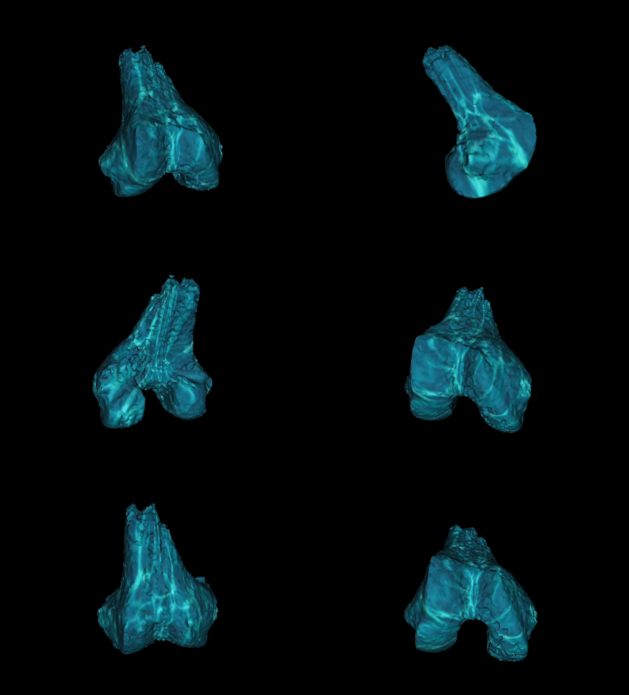
Osteoarthritis (OA) is a disease of the entire joint, involving whole joint tissue change. The bone-cartilage interface is a complex functional unit and bio-composite at the center of joint function and disease in which the individual components interact cooperatively and synergistically. The causes of degenerative changes in knee OA(KOA) are complex and involve interrelated biological, mechanical, and structural pathways. While KOA is a complex disease with multiple phenotypes that can influence its initiation and progression, mechanical stress has become a primary consideration in assessing the nature of the disease. Magnetic resonance imaging (MRI) allows the detection of the whole spectrum of pathological joint tissue changes. Clinically, MRI revealed Bone Marrow Lesions (BMLs) are occasionally found in KOA patients, preceding the onset of articular cartilage degeneration. BMLs are indicators of KOA progression and an important risk factor for structural deterioration. In this project, we aimed to demonstrate the relationship between kinematic features and quantitative analysis of bone lesions in pre/early knee OA based on MRI images.


